Vocabulary
|
|
|
The reproduction
The female reproductive apparatus:
-Ovaries: Are two orfans the size an almond
-The fallopian tube: these are two channels which join the ovaries to the uterus
-Uterus: This is a cavity which is prepared for a fertlised ovum and for housing the embryo
-Vagina: this is a flexible channel wich comunicates the uterus with the exterior
The male reproductive apparatus:
-Testicles: these are two organs that make the spermatozoids
-Epididymis: These are structures located above the testicles
-Vas deferens: small channels through which the spermatozoids travel to the seminal vesicles
-Seminal vesicles: the spermatozoids are stored here
-Urethra: is the tube than runs from the bladder and seminal vesicles
-Penis: is to deposit the spermatozoids inside the vagina
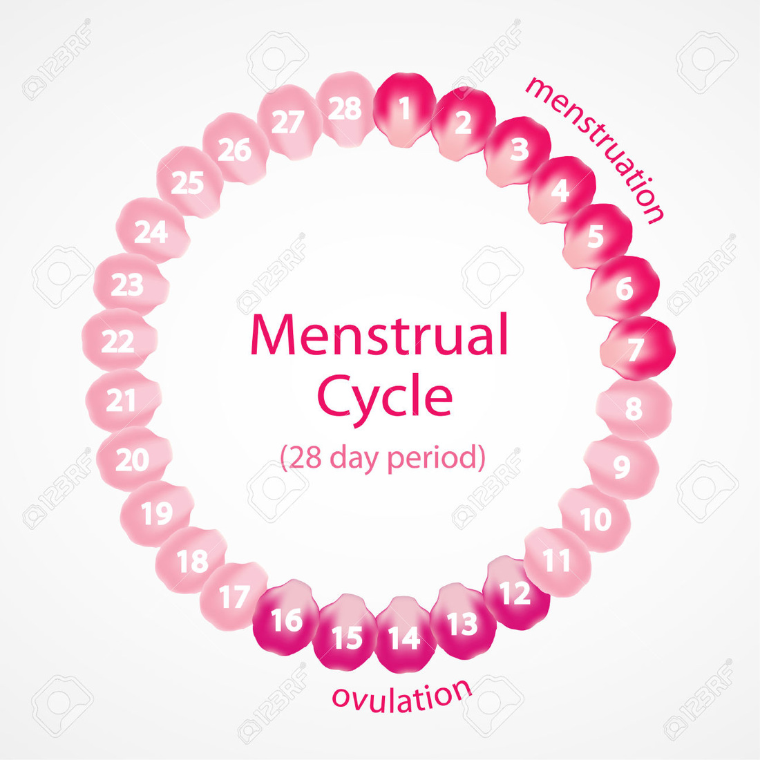
The menstrual cycle
During the marutity ovum, the uterus prepares itself for a possible pregnancy, as this is where the fertilised ovum would be installed, and covers the interior with mucous which is in contact with a great namber of blood cells
This layer is known as endometry.
When the ovum is mature, it leaves the ovary and passes along the fallopian tube. From this moment it has a life span of 24 hours
The endometry is released 14 days after the death of the ovum. This is how mnstruation occurs, which is a loss of blood through the vagina which lasts berween 4 and 5 days
Fertilisation, pregnancy and birth
Fertilisation is the union berween an ovum and a spermatozoid to form an egg cell. This process is produced in the Fallopian tube .
The spermatozoids are introduced into the vagina during coitus. It the spermatozoid is unable to find an ovum in the Fallopian tubes, it will die after about four or five days. It the spermatozoids find an ovum they surround it and try to penetrate it only one will archieve this.
Once the fertilisation has occurred in the Fallopian tube the zygote begins to divide many times during its route to the uterus. The installation of the embryo on the walls of the placenta is known as nesting and it occurs some 7 days after fertilisation.
The embryo is surrounded by a membrane known as the chorion. In the area of contact between the chorion and the endometry, the chorion grows some prolongations which penetrate the endometry.
The embryo and later the foetus recibe oxygen and nutrients necessary for growth along the umbilical cord
The birth occurs at the end of the pregnancy, there are three stages:
- Dilatation of the uterine neck: this begins with the contractions of the walls of the uterus and the dilatation of the neck of the uterus.
- Expulsion of the foetus: the contractions of the walls of the uterus and the pressure of the abdominal muscles push the foetus along the vagina to the exterior
- Expulsion of the placenta: Later the placenta is released from the walls of the uterus and is expelled together with the other remains
SENS
EYE:
Semi-circular canals are curved tubes filled with a liquid which moves when you move. Nerve endings in the canal walls carry information about the fluid´s movement to the brain
HEARING
Sound waves are collected by the funnel-shaped ipinna or each ear, and pass down a short tube to the ear drum. The ear drum is a thin sheet of skin, and sound waves make ir vibrate.
When the ear drumvibrates, the ear ossicles move against each other in such a way that they lever the oval window in and out. This causes vibrations to pass along a tube called the cochlea, this transformed the sounds into the nerve impulses which give the sensation of sound
- An eye is like a camera.
- Cornea: this is the transparent window at the front of an eyeball
- Iris: this is the coloured part of the eye
- Lens: is held in place by suspensory ligaments
- Pupil: gets bigger in dim light and gets smaller in bright light
- Focucusin muscles: hold the lens in place
- Retina: a layer o light-sensitive cells
- Optic nerve: connecting an eye with the brain
Semi-circular canals are curved tubes filled with a liquid which moves when you move. Nerve endings in the canal walls carry information about the fluid´s movement to the brain
HEARING
Sound waves are collected by the funnel-shaped ipinna or each ear, and pass down a short tube to the ear drum. The ear drum is a thin sheet of skin, and sound waves make ir vibrate.
When the ear drumvibrates, the ear ossicles move against each other in such a way that they lever the oval window in and out. This causes vibrations to pass along a tube called the cochlea, this transformed the sounds into the nerve impulses which give the sensation of sound
NERVOUS SYSTEM
The nervous system has two parts
- The central nervous system is formed by the encephalon and the spinal cord. These organs are inside the cranium and the spinal colum, the nervous tissue is isolated from the bone by three membranes known as meninges
- The peripheral nervous system consists of nerves. These are made un of the axons of neurons. In the same way that there are sensory and motor neurons. According to their origin, the nerves can be classified as cranial and spinal
endocrine system
Is formed for a glands, these produced diferent hormones:
- Thyroid: thyroxine, it activates the cellular metabolism
- Parathyroid: parathormone, it increases the level of calcium in the blood
- Suprarenal glands: adrenaline, it favours intense muscular activity
- Pancreas: insulin, it decreases the level of glucose
- Ovaries: estrogens, the appearance of female sexual characteristics
- Testicles: testosterone, the appearance of masculine sexual characteristics
- Hypophysis: prolactin, it favours the secretion of milk in the breast after giving birth
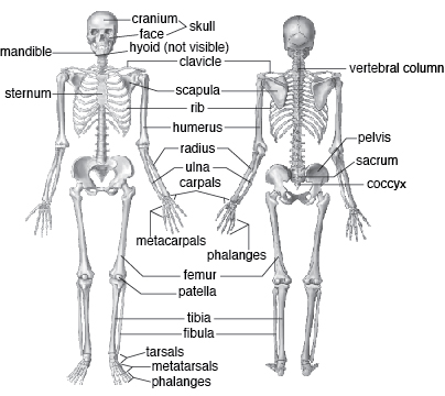
THE SKELETON
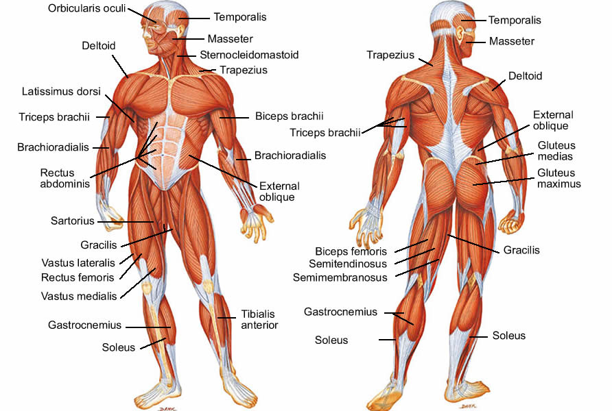
MUSCLES